What is keratoconus and what are the treatment options?
What is keratoconus?
Keratoconus is a disorder that affects the cornea - the clear window on the front of the eye. The cornea functions to focus light into your eye to enable clear vision. Keratoconus typically involves both eyes but is usually asymmetric with one eye worse than the other. There is often a history of frequent eyeglass prescription updates that fail to fully correct the vision.

Keratoconus is associated with a change in the shape of the cornea that causes blurry vision.
The cornea assumes a subtle cone shape in keratoconus.
Early diagnosis, close monitoring and early corneal crosslinking are the key steps to prevent vision loss associated with keratoconus
Without prompt treatment and monitoring, keratoconus typically progresses and causes an ongoing decline in vision over time. As keratoconus progresses, the cornea thins and steepens causing worsening shortsightedness and irregular astigmatism. As the shape of the cornea changes with keratoconus, the ability of the eye to clearly focus light is lost and vision becomes blurry and distorted. Daily tasks that we all take for granted like reading or driving can become difficult or impossible. In mild keratoconus, blurry vision may improve with glasses. In more advanced cases, rigid contact lenses and/or surgery may be required to maintain vision.
What causes keratoconus?
Despite a large amount of research, the exact cause of keratoconus remains unknown. What we do know is that there is a strong association with atopic (allergic) disorders such as asthma, eczema and hayfever and many people with keratoconus have a significant history of eye rubbing. A small number of patients will have a family history of keratoconus, in most cases, however, keratoconus is not inherited.

Keratoconus has a strong association with eye rubbing
Onset is typically in the teenage years but may occur earlier in childhood or later in life. Early onset is often associated with more aggressive keratoconus whereas late onset can often be a milder form.
Risk factors for developing keratoconus
Keratoconus is more common than previously thought. Up to one in two hundred adolescents in New Zealand may have keratoconus and up to one in forty-five for those of Māori descent. There are several risk factors that are associated with an increased chance of developing keratoconus.
Atopy (asthma, eczema, hayfever)
Eye rubbing (very strong association)
Connective tissue disorders
Family history of keratoconus
Retinitis pigmentosa
Down syndrome
Floppy eyelid syndrome
Sleep apnoea
Ethnicity (keratoconus is more common in certain ethnic groups such as Māori and Pasifika)
Diagnosis of keratoconus
Very early keratoconus can be difficult to detect and many people may be unaware that their vision is not normal or that there may be a problem with their cornea. In some cases, there may be a history of worsening vision requiring frequent updates for the prescription of glasses.
The definitive diagnosis of keratoconus is based on a clinical examination and high-resolution scanning of the cornea (corneal tomography) to evaluate the precise shape and thickness. Keratoconus has certain features that can be detected on the corneal tomography scan. These features will be visible in many cases before the vision begins to deteriorate.
Diagnostic procedures
Slit lamp examination
Retinoscopy
Corneal tomography scanning
Treatment for keratoconus
The aim of treatment for keratoconus is to halt changes in the corneal shape and maintain vision. The gold standard treatment for progressive keratoconus is corneal collagen crosslinking (also known as corneal crosslinking). This treatment is a surgical procedure that is designed to strengthen the cornea and halt the further progression of keratoconus along with any associated loss of vision. Corneal crosslinking is an effective treatment that has a high rate of success and rapid recovery.
You can find out more about corneal crosslinking here. It is also important to avoid eye rubbing as this may make your keratoconus worse. Many people with keratoconus have itchy and sensitive eyes. There are several eye drops that can help reduce itchiness and irritation to help break the cycle of eye rubbing.
Once the keratoconus has been stabilised, it is then possible to look at strategies to restore vision.
Restoring vision in keratoconus
Improving vision often requires glasses, contact lenses or surgery. It is important to understand that glasses or contact lenses may help improve vision but are not effective treatments for halting the progression of keratoconus. The only effective treatment that has been proven to halt the progression of keratoconus is corneal crosslinking.
Glasses and contact lenses are not a treatment for keratoconus and will not stop progression and worsening of vision
For people with mild keratoconus, glasses may still work well to improve vision. In more severe cases of keratoconus, glasses are often ineffective and fail to provide clear vision. In these advanced cases where the cornea often has an irregular shape, contact lenses may be a good option for vision correction. Contact lens fitting is keratoconus is a specialist skill and, in many cases, a custom-fitted rigid gas permeable (RGP) contact lens is required to achieve clear vision. Although there are many different types of RGP lenses, there are some people who are intolerant of contact lenses or who would rather have a surgical solution to permanently correct their vision.
Surgical options to restore vision in keratoconus
There are several surgical options that are available for vision correction in people who have keratoconus.
Non-surgical options (glasses, and/or contact lenses) to correct vision are usually attempted before considering surgery.
Corneal crosslinking may provide a modest improvement in vision without glasses or contact lenses, however, in most cases, this will not be sufficient to provide clear vision and other surgery may be required.
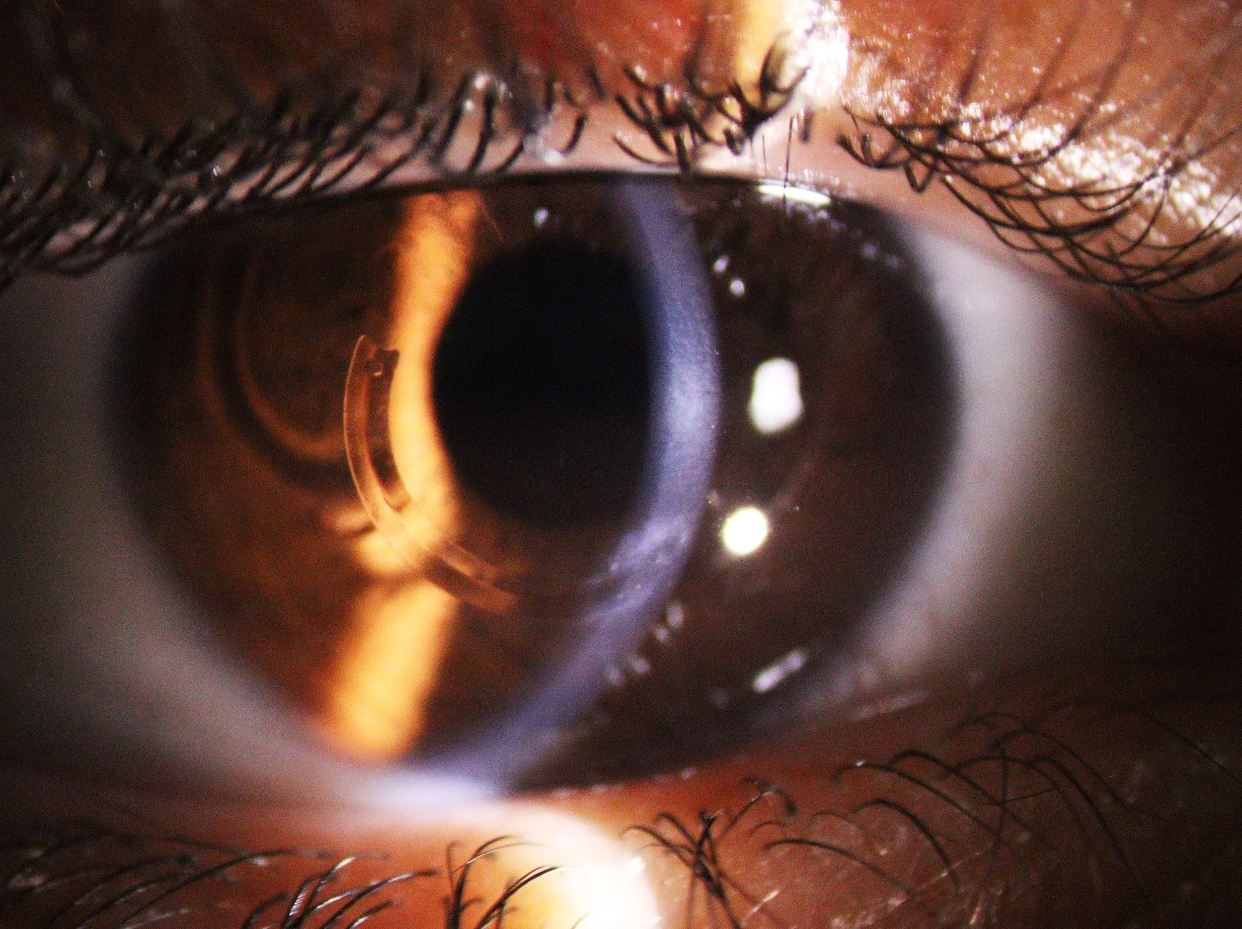
Intracorneal ring segments (ICRS) provide a good improvement in vision for approximately 80% of patients. This is a reversible procedure that involves implanting a thin transparent inlay into the cornea to improve the shape. This can be a good option for people who cannot tolerate contact lenses. The improvement in shape after implanting ICRS may mean that a contact lens or glasses can be effective to improve vision.
In some cases, including those who have ICRS and do not want to wear glasses or contact lenses, it may be possible to insert an Implanted Collamer lens (ICL) to permanently correct your vision.
What should I do if I have keratoconus?
If you have keratoconus or have risk factors for the development of keratoconus, you will need a complete eye examination including corneal tomography. It is essential to complete a baseline tomography corneal scan to accurately assess and document any progression in the future. Please call for an appointment so we can arrange an assessment and imaging. Your appointment is the perfect time to answer any questions you may have including arranging crosslinking or other vision-saving/restoring treatment.
You can book an appointment online or call Gaynor on ☎ 0800 CATARACT (0800 228 27 228) or (07) 834 6335. If you need an appointment to discuss keratoconus, cross-linking, or vision-correction surgery please get in touch and we will do our best to arrange an appointment within the next 1-2 weeks.
For any other questions please feel free to email info@jamesandeye.co.nz